Please read the important information regarding these games at the bottom of the page.
The electrocardiogram – looking at the heart of electricity
A milestone in heart diagnosis
The electrocardiogram or ECG (sometimes called EKG) is today used worldwide as a relatively simple way of diagnosing heart conditions. An electrocardiogram is a recording of the small electric waves being generated during heart activity.
The electric currents in the heart have been measured for more than a hundred years, but the fundamental function of the ECG as we know it today was developed by the Dutch scientist Willem Einthoven in the beginning of the 20th century. In 1924 Einthoven was awarded the Nobel prize in Physiology or Medicine "... for his discovery of the mechanism of the electrocardiogram."
What makes the heart beat?
|
|
The electric activity starts at the top of the heart and spreads down. |
A normal heart beat is initiated by a small pulse of electric current. This tiny electric "shock" spreads rapidly in the heart and makes the heart muscle contract.
If the whole heart muscle contracted at the same time, there would be no pumping effect. Therefore the electric activity starts at the top of the heart and spreads down, and then up again, causing the heart muscle to contract in an optimal way for pumping blood.
Where does the electricity come from?
In the heart there are cells specialized in producing electricity. These are called pacemaker cells. They produce electricity by quickly changing their electrical charge from positive to negative and back.
The first electric wave in a heart beat is initiated at the top of the heart. Because of the heart muscle cell's ability to "spread" its electric charge to adjacent heart muscle cells, this initial wave will be enough to start a chain reaction.
The challenge of registering millivolts
Making the electrodes sensitive enough was a challenge in the early days of the ECG. In the late 1800's early attempts to measure the electric activity in frog's hearts were successful only when the hearts were exposed directly to the measuring equipment. The measuring conditions were indeed difficult; the scientists wanted to be able to measure the electric signals without having to enter inside the body. The problem was that the electric wave got weaker since it had to travel through bone and body tissue before reaching an electrode applied on the skin. This problem was solved a couple of decades later by Willem Einthoven. He managed to improve the sensitivity of the ECG by using a string galvanometer.
 |
| Willem Einthoven |
A lot of Einthoven's terminology is still being used and his original research remains fundamental to electrocardiography today.
From electrode to paper
The electric waves in the heart are recorded in millivolts by the electrocardiograph. The waves are registered by electrodes placed on certain parts of the body. Each electrode controls an ink needle that writes on a grid paper. The higher the intensity of the electric wave, the higher up the needle will move on the paper. The paper moves at a certain speed beneath the needle, resulting in an ink curve.
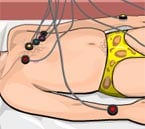 |
| Sensitive electrodes are placed on certain parts of the body. |
The leads & Einthoven's triangle
The electrodes are typically twelve in number. The stretch between two limb (arm or leg) electrodes is called a lead. Einthoven named the leads between the three limb electrodes "standard lead I, II and III" referring to the two arm electrodes and the left leg electrode. He studied the relationship between these electrodes, forming a triangle where the heart electrically constitutes the null point. The relationship between the standard leads is called Einthoven's triangle. Einthoven's triangle is used when determining the electrical axis of the heart.
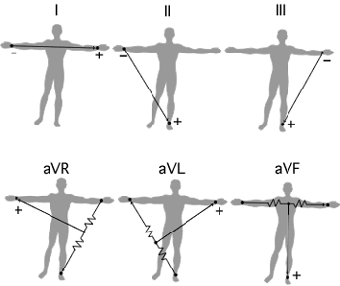 |
| The standard leads (top) and
the augmented leads (bottom) reflect the limb
electrodes (left arm, right arm, left leg) used
to record the heart's electrical axis in the
frontal plane. Illustration by Urban Frank. |
Mountains & valleys
An ECG curve has different characteristics depending on the location of the electrode recording it. When the curve falls below the base line it shows a negative deflection and when it rises above the base line it is a result of positive deflection. A negative deflection indicates that the recorded wave has traveled away from the electrode and a positive deflection means it has traveled towards it.
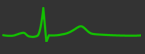 |
| An ECG curve reflects the perspective of the electrode recording it. |
Abnormal heartbeats & conduction defects
If the electric or muscular function of the heart is disturbed for some reason, it will affect how the electric signals spread through the heart muscle. One example is arrythmia, a condition where the heart beats irregularly due to a defect in the electric conduction system.
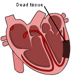
A cardiac infarction is another condition which results in dead tissue in a part of the heart muscle, and therefore the electric signal cannot travel through that area.
A left or right bundle branch block delays the electric wave from spreading to the left or right part of the heart. Sometimes these conditions affect the heart's ability to pump blood.
 |
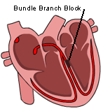 |
| Cardiac infarction | Left bundle branch block |
Cells directing the heart muscle
When the heart muscle is at rest, the pacemaker cells are negatively charged and when the heart contracts they are positively charged. When a positive wave is recorded by a positive electrode, the ECG curve will be pointing upwards and vice versa.
Repolarization and depolarization
The cells change their electric charge by means of depolarization and repolarization. Depolarization occurs when negatively charged ions inside the cell travel out from the cell through the cell membrane and positively charged ions travel in (repolarization).
Assisting the heart
 |
| A pacemaker's job is to deliver a steady and regular electric impulse to the heart. |
Pacemaker is also the name of a device that can help stabilize the electric conduction system when the heart's electric function is not working properly. The pacemaker is surgically placed in the chest, and delivers a steady rhythm of "starting" waves where this function is defective.
Play the Electrocardiogram Game
More about the discovery of the electrocardiogram and Nobel Laureate Willem Einthoven
First published: August 2002
MLA style: "The electrocardiogram, ECG". Nobelprize.org. Nobel Prize Outreach AB 2022. <http://nobel-external-educationalgames-app.azurewebsites.net/educational/medicine/ecg/ecg-readmore.php>