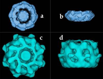
Electron Microscopy: Nuclear Pore ComplexA three-dimensional image of the nuclear pore complex (NPC), revealed by electron microscopy. A-B The NPC in yeast. Figure A shows the NPC seen from the cytoplasm while figure B displays a side view. C-D The NPC in vertebrate (Xenopus). Figure A shows the NPC seen from the cytoplasm while figure B displays a side view. |
 |
|
Images kindly provided by Dr Christopher W. Akey, Department of Biophysics, Boston University. Reference: Three-Dimensional Architecture of the Isolated Yeast Nuclear Pore Complex: Functional and Evolutionary Implications, Qing Yang, Michael P. Rout and Christopher W. Akey. Molecular Cell, 1:223-234, 1998.
|